Bone Analysis
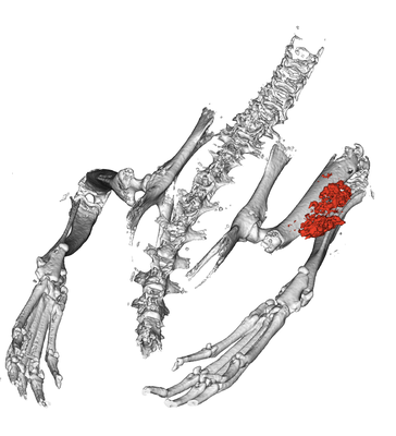
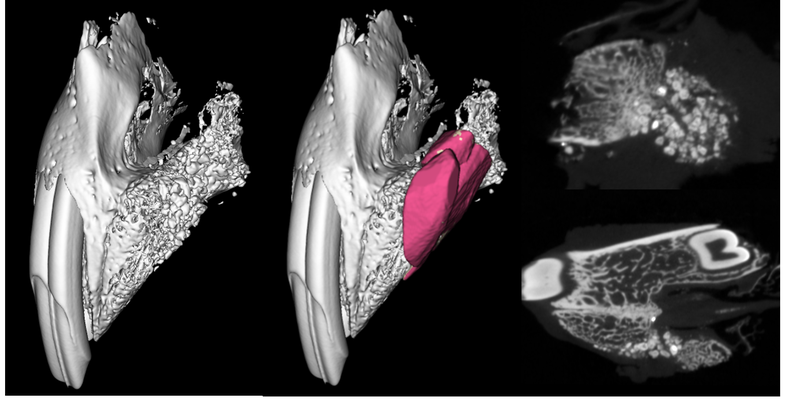
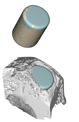
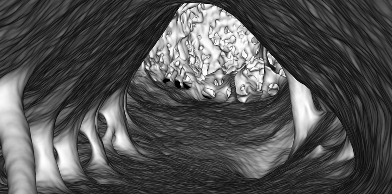
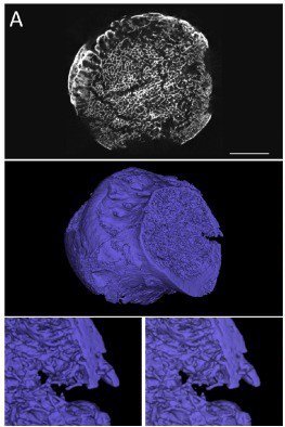
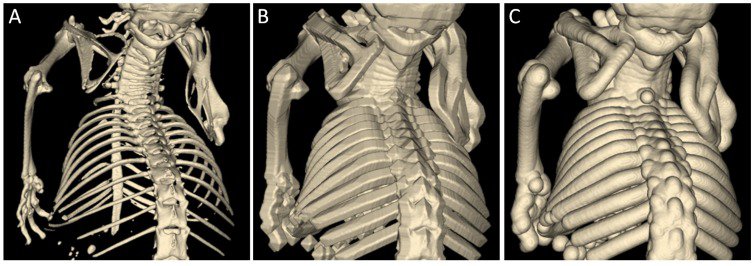
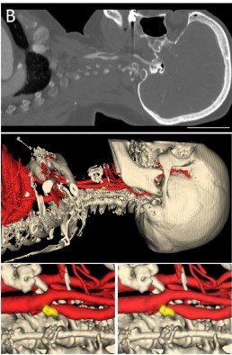
The analysis of different bone structures and features, the evaluation and quantification of new bone formation as well as the occurrence of calcification were integrated into the software. Various studies on different bone filling materials have been successfully performed. Also, human bone image data have been successfully reconstructed and analyzed. The representative images show the alveolar cleft on a New Zealand White rabbit skull, bone segmentation via thresholding using box kernel (B) and spherical kernel, the head of a human femur (A-blue), bone segmentation and labeling (C).
